


The Light of the Titans: How a Laser Breakthrough is Revolutionizing Cellular Imaging

For nearly a century, the field of microscopy has been defined by a constant struggle against the inherent transparency of life. Biological cells, largely composed of water, are notoriously difficult to visualize; they lack the density required to scatter light, rendering their internal architecture—the complex machinery of mitochondria, nuclei, and proteins—nearly invisible under standard illumination.
In 1930, Dutch physicist Frits Zernike solved this by inventing phase-contrast microscopy, a technique that transformed subtle shifts in light waves into stark, visible differences in brightness. This innovation earned him a Nobel Prize in 1953 and opened the door to modern cell biology. Today, a team of physicists at the University of California, Berkeley, led by Professor Holger Müller, has effectively brought this "phase-contrast" revolution into the atomic age. By developing an ultra-intense laser phase plate for electron microscopy, the team has created a tool capable of imaging biological structures previously thought too small to be seen.
The Technological Leap: From Light to Electrons
Electron microscopy (EM) has long been the gold standard for high-resolution imaging, capable of magnifying objects up to 10,000 times more effectively than traditional light microscopes. Specifically, cryo-electron microscopy (cryo-EM)—which involves freezing samples to protect them from radiation damage—has revolutionized drug discovery by allowing scientists to map the 3D structures of proteins.
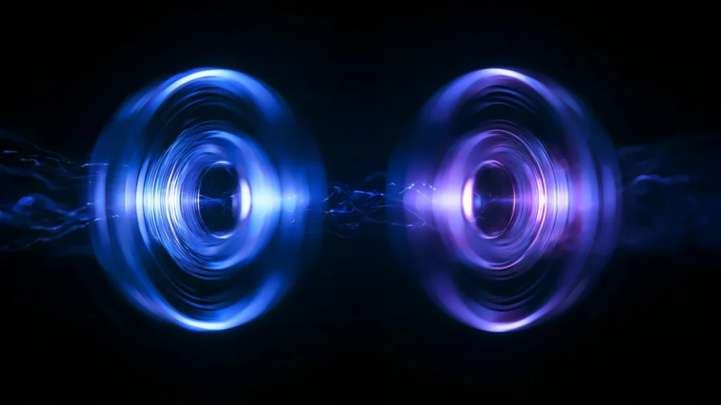
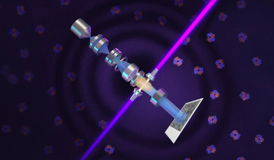
However, cryo-EM has a "size ceiling." Most human proteins are remarkably small, and current technology often fails to capture the clear, high-contrast images required to analyze them. Enter the "Theia" microscope, named after the Greek Titaness of light and radiance. By integrating an incredibly powerful continuous-wave laser directly into the electron beam path, Müller’s team has created a system that alters the phase of electrons, dramatically boosting image contrast without destroying the delicate specimen.
A Chronology of Innovation
The path to this breakthrough was neither short nor simple. It is a story of a 15-year quest to harness light in a way that had never been done before.
The 2010 Proposal
The foundational idea was born in 2010 when Holger Müller and UC Berkeley professor emeritus Robert Glaeser proposed using a high-intensity laser to manipulate electron beams. Their hypothesis was simple but technically daunting: if one could force a laser to interact directly with an electron beam, one could induce a phase shift that would enhance contrast without the need for traditional, damaging staining techniques.
A Decade of Engineering
For the next ten years, the team faced a primary obstacle: the sheer physics of the required laser intensity. To shift the phase of an electron beam by 90 degrees, they needed a laser more powerful than anything used in standard laboratory settings. They eventually engineered a spherical, mirror-lined cavity that forced a laser beam to reflect over 10,000 times. The result was a focal point of 75 kilowatts concentrated into a few microns—a intensity surpassing that of industrial welding lasers or even military-grade directed-energy systems.
The Modern Implementation
With support from Biohub, the researchers successfully installed this laser system into a state-of-the-art Thermo Fisher Krios cryo-EM instrument. In recent tests, they confirmed that the laser provides a "step-function" improvement in visibility, particularly for the most challenging, small-scale biological samples.
The Challenge of the "Invisible" Proteome
To understand the scale of this achievement, one must look at the "proteome"—the entire set of proteins expressed by the human body. Proteins are measured in daltons, and the vast majority of human proteins are under 70 kilodaltons (kDa).
Standard cryo-EM methods struggle significantly with anything below this threshold. Because these proteins are so small, they scatter very few electrons, resulting in "noisy" images that make structural analysis nearly impossible. By utilizing the laser phase plate, the Berkeley team has successfully imaged structures as small as 50 kDa, with the theoretical potential to reach 17 kDa—the size of myoglobin.
"The bottom line is," notes Professor Müller, "if you have a large protein and a really good sample, you may not need the phase plate. But for a small protein and a difficult sample, laser-on is best. This fills an enormous gap in our knowledge."
Implications for Cryo-Electron Tomography (Cryo-ET)
While the improvement to cryo-EM is significant, the impact on cryo-electron tomography (cryo-ET) may be even more transformative. Unlike cryo-EM, which often looks at isolated proteins in a solution, cryo-ET captures images from multiple angles to construct a 3D view of the cell in its natural state.
Bridget Carragher, the founding technical director of imaging at Biohub, provides a vivid analogy for the current state of the field: "We’re looking at small, very complicated cellular material that’s incredibly crowded inside the cell. It’s like a forest of trees, and you’re trying to find one leaf on one tree in there. Cryo-ET needs a dramatic step forward in contrast, so we can start to see what’s going on inside the cell. That’s what the laser phase plate promises."
By allowing researchers to see how proteins interact within the crowded, chaotic environment of a living cell rather than in isolation, the technology moves us closer to a "systems biology" understanding of health and disease.
Official Perspectives and Future Directions
The scientific community has reacted with cautious optimism, acknowledging that this is a rare "jump" in capability. The collaboration between UC Berkeley and Biohub, along with partnerships with instrument manufacturers like Thermo Fisher Scientific, signals a clear path toward commercializing this technology.
The Biohub Approach
At Biohub’s facility in Redwood City, Carragher’s team is building a second-generation system that uses two laser beams operating at right angles. By splitting the power, the researchers minimize the risk of hardware burnout and reduce optical distortions, pushing the resolution limits even further.
A New Era for Medicine
Stephani Otte, Biohub’s Vice President of Imaging Science, believes the implications for medicine are profound. "We are going to be able to see how molecular machines operate inside the living cell, in context, for the first time," Otte states. "What was once invisible will become visible—and that changes everything about how we understand disease."
If scientists can map the interactions of the 90% of proteins that were previously too small to visualize, it could lead to an explosion in drug discovery. Many of the most elusive targets for cancer and neurodegenerative diseases are small proteins that current imaging technology simply cannot resolve.
Conclusion: Bridging the Gap
The work of Müller, Glaeser, and their collaborators—including postdoctoral fellows Petar Petrov and Jessie Zhang—represents a convergence of high-energy physics and cellular biology. By revisiting the principles that earned Frits Zernike a Nobel Prize in 1953 and applying them to the 21st-century world of electron beams, they have bridged the gap between what we can hypothesize and what we can actually see.
As the team refines the technique, the focus will shift to using a tightly focused electron beam, which could double the signal-to-noise ratio once again. With these advancements, the "invisible" machinery of human life is finally coming into focus, promising a future where the inner workings of our cells are no longer a mystery, but a map for the next generation of medical breakthroughs. The Titaness Theia has indeed brought light to the darkest corners of the biological world.